Continually Updated Clinical Reference
 
 
  All Sources     eMedicine     Medscape     Drug Reference     MEDLINE
 
eMedicine - External Auditory Canal Atresia : Article by

Quick Find
Authors & Editors
Introduction
Clinical
Differentials
Workup
Treatment
Follow-up
Miscellaneous
Multimedia
References

Related Articles
Cleft Lip and Palate

Crouzon Syndrome

Microtia

Osteogenesis Imperfecta


Patient Education
Click here for patient education.


Author: Rahul K Shah, MD, FAAP, Assistant Professor of Otolaryngology and Pediatrics, George Washington University, Children's National Medical Center; Attending Physician, Department of Otolaryngologoy, Children's National Medical Center

Rahul K Shah is a member of the following medical societies: Alpha Omega Alpha, American College of Surgeons, Massachusetts Medical Society, Phi Beta Kappa, Society of University Otolaryngologists-Head and Neck Surgeons, and Triological Society

Coauthor(s): Udayan K Shah, MD, Attending Surgeon, Division of Otolaryngology, Alfred I duPont Hospital for Children

Editors: Orval Brown, MD, Director of Otolaryngology Clinic, Professor, Department of Otolaryngology-Head and Neck Surgery, University of Texas Southwestern Medical Center at Dallas; Mary L Windle, PharmD, Adjunct Assistant Professor, University of Nebraska Medical Center College of Pharmacy, Pharmacy Editor, eMedicine.com, Inc; John E McClay, MD, Assistant Professor, Department of Otolaryngology, Division of Pediatric Otolaryngology, Children's Medical Center, University of Texas Southwestern Medical School; Paul D Petry, DO, FACOP, FAAP, Consulting Staff, Freeman Pediatric Care, Freeman Health System; Maureen Strafford, MD, Arnold P Gold Foundation Associate Professor, Departments of Anesthesiology and Pediatrics, Tufts University and Tufts-New England Medical Center

Author and Editor Disclosure

Synonyms and related keywords: external auditory canal atresia, congenital aural atresia, congenital atresia of the external auditory canal, EAC, EACA, EAC atresia, conductive hearing loss, ossicular malformations, craniofacial syndromes, atretic external canal, microtia, otitis media, OM, mastoiditis, facial nerve palsy, cholesteatoma

Background

External auditory canal atresia (EACA) is a rare congenital disorder. Accurate counseling and referral for these children and their families demands an appreciation of the range of management options and indications for intervention. This article reviews the history, etiology, and epidemiology of EACA, as well as the clinical care of children with this condition.

EACA was first described in Western medicine by Paulus of Aegina in the seventh century AD. Paulus recommended simple incision for opening the atresia. Later, physicians used a hot iron probe to maintain canal patency after incising the atresia. The first operation to correct an atresia of the external auditory canal (EAC) was performed in 1882 by Kiesselbach. Unfortunately, the operation left the child with a facial nerve paralysis. In 1914, Page reported on a series of 8 patients who underwent operation. Of these 8 patients, 5 had a subjective improvement in their hearing after surgery.

In the early 20th century, techniques for correction of the atretic external canal have involved opening the antrum and aditus of the ear and lining the cavities with a skin graft. In these surgeries, the atretic bony plate was untouched, and the resulting hearing improvement was poor. In the past 50 years, improved radiologic and audiologic assessments coupled with improvements in surgical technology, such as the operating microscope and facial nerve monitoring, have resulted in good surgical success rates for properly chosen patients with EACA.

The future of medical and surgical interventions for patients with EACA is exciting and includes such novel approaches as computer-aided surgery as well as critical reviews of past surgical results, with further attempts to increase success rates and minimize morbidity.

Pathophysiology

The EAC is comprised of medial bony and lateral cartilaginous portions. The bony component changes from half of the length of the tubular EAC in children to two thirds of the length of the tubular EAC in adults.

The lateral soft tissue portion of the EAC begins to form at 26-28 weeks' gestation, when the epithelial plug arising from the first branchial cleft begins to canalize. During the sixth month of development, the medial bony portion is created out of the temporal bone's mesenchymal condensation. As canalization occurs, the mastoid separates from the mandible and grows posteriorly and inferiorly. The posteroinferior growth of the mastoid brings the facial nerve from the middle ear and brings the facial nerve and middle ear into its position at birth (ie, posterior and inferior to the EAC).

Congential atresia of the EAC is caused by a failure of canalization of the epithelial plug portion of the first branchial cleft. Persistence of the tympanic ring results in a bony atresia plate at the level of the tympanic membrane. Ossicular malformations may be seen as they arise from the first branchial cartilage (ie, Meckel cartilage).

Failure of the EAC to canalize means that sound cannot reach the tympanic membrane; thus, a conductive hearing loss results (see Media files 2-3). Concomitant ossicular malformations may result in additional conductive hearing loss. In addition, 11-47% of patients also have a sensorineural hearing loss in the affected ear.
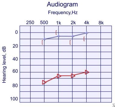
EACA may be seen with microtia (ie, maldevelopment of the external ear). The auricle develops from ectodermal condensations and mesodermal condensations (referred to as the hillocks of His) at 5 weeks' gestation.

Case reports have detailed duplication anomalies of the first branchial cleft occurring in conjunction with a congenital aural atresia. The implication for the evaluating physician is to keep in mind that EACA and duplication anomalies can be present in the same patient, and both may need to be simultaneously addressed (surgically).

Frequency

United States

According to Jahrsdoerfer, the incidence of ear malformations in New York City from 1952-1962 was 1 in 5800 births.1 In one year of this study, the incidence of EACA was 1 in 8000 births.

International

EACA is rare in persons with a normal pinna. The incidence of EACA with concurrent microtia is 1 in 10,000-20,000 live births. Unilateral EACA is 3-6 times more likely to occur than bilateral atresia. The right ear is more often involved than the left ear. Family histories positive for EACA are noted in 14% of patients.

Mortality/Morbidity

Intervention to treat bilateral EACA is important for hearing and language development. Social stigma may be experienced because of cosmetic or developmental issues, especially as grade school begins.

  • Otitis media (OM) is a common pediatric diagnosis that is difficult to identify and manage in children with EACA. EACA complicates the diagnosis of OM. Symptoms of pain, fever, irritability, and worsening hearing loss or imbalance warrant evaluation. The contralateral ear may also show otitis. Oral antibiotics may be started presumptively. Although CT scanning is not a routine screening test for these children, it reveals soft tissue density in the middle ear cleft. Clinical management of OM is not significantly different, and complications of mastoiditis, facial nerve palsy, and intracranial complications must be monitored.
  • Once complications have been encountered, CT scanning to examine the interrelationships of the otologic structures is certainly warranted. OM refractory to oral antibiotics may require insertion of tympanostomy tubes. However, these ventilation tubes should only be inserted in the normal ear and not the affected contralateral ear. The need for middle-ear ventilation to treat OM may prompt earlier correction of EACA as the diagnosis is made earlier. Recurrent OM is an age-limited problem that resolves with time.
  • Canal cholesteatoma (epithelial inclusion cyst) may occur medial to the atretic ear canal. Canal cholesteatomas may be identified radiographically before symptoms begin because of widening of the EAC. Patients with infection of the cholesteatoma present with pain and fever. Treatment is primarily surgical with adjunctive use of oral or systemic antibiotics.
  • Traditionally, unilateral EACA was not believed to limit children's language. Recently, therapies for hearing loss have appreciated the importance of bilateral hearing for optimal speech and language development. In addition to long-term development, bilateral hearing is important for hearing in a noisy environment (eg, in groups, with background noise) and for sound localization (eg, knowing from which side a car is approaching).

Sex

EACA occurs more often in males than in females.

Age

EACA occurs with a normal pinna or with an abnormal pinna (ie, concomitant microtia). When the atresia occurs in the setting of a normal pinna, the average age of diagnosis is 2.5 years. However, when EACA occurs with abnormal development of the pinna, the average age of diagnosis is 3.5 years.


History

Diagnosis of a congenital ear malformation is usually made at birth, when a malformed pinna or atretic canal is noticed during the secondary survey of the newborn. Some cases may not be noticed at birth, such as those with a normal pinna and a blunted or partially patent canal. Some of these patients are detected during a screening examination performed in school.

  • When soliciting information regarding the history of the present illness, determine how and when the problem was discovered and ask about the child's language and social development and associated problems.
  • Survey the family for history of hearing loss and syndromic conditions and for genetic information.
  • Inquire into maternal and neonatal health.
  • Identify the state of patient's social and language development.
    • The child's interactions with other children, caregivers, and family are important.
    • The patient's language development, with age of babbling and first words or number of words, is a useful measure.
    • Children with unilateral external auditory canal atresia (EACA) usually have normal speech and language development. Hearing and learning may be impeded in noisy environments, such as group discussions or large lectures, and sound localization may be a problem at play and when outdoors.
  • Rarely, an adult presents with a report of a hearing loss, and the physical examination reveals EACA.
  • Reports of vertigo may require a formal assessment of the vestibular system.
  • Pain, fever, or drainage from the atretic ear may indicate cholesteatoma or infection of the ear canal.
  • History of facial nerve problems must be identified, particularly before surgical repair. Problems with eye closure may manifest as dry, itchy, or sandy eyes and rarely as exposure keratitis. Problems drinking liquids or drinking with a straw may also indicate facial nerve dysfunction.

Physical

The global level of functioning of the child should be addressed first. Complete head and neck examination then follows with attention to craniofacial anomalies. Then, otologic structures are assessed. For complete care of patients with EACA, physical examination must be combined with radiographic evaluation.

Examine the remainder of the body so as not to overlook other abnormalities, especially cardiac, renal, or ophthalmologic malformations.

  • Pinna: Assess for microtia.
  • Concha: A thumbprint concha is a classic sign for EACA (see Media file 1). The meatus of the EAC may simply be a pinpoint, indicating severe canal stenosis rather than complete atresia. Care of these patients is similar to that of patients with EACA.
  • Congential ear malformations
    • These can be divided into major and minor forms.
    • Major ear malformations consist of an absence of the external canal and tympanic membrane, a small middle-ear cavity, and malformations of the malleus and incus. Microtia and a hypoplastic mandible are concomitant findings in patients with major ear malformations.
    • Minor ear malformations consist of abnormal ossicles with a patent EAC and tympanic membrane, although the latter 2 structures may be smaller than normal.
  • Lateral skull
    • The relationship of the temporomandibular joint in relation to the mastoid tip and middle ear is documented.
    • Mastoid tip development should be assessed and especially compared to the contralateral side. Poor mastoid tip development portends abnormal facial nerve anatomy.
  • Facial nerve
    • Evaluation should be performed using scoring or grading according to established systems, such as those by Fisch or House and Brackmann.
    • An objective assessment may be indicated in cases of palsy (see Lab Studies).
  • Documentation
    • Photographic records of anomalies aid referrals, teaching, and objective analysis of surgical results.
    • Images should be standardized with respect to lighting and dimensions.
    • Appropriate consents from the family and patient should be obtained for teaching and publication purposes, ideally at the time of such documentation.

Causes

Precise etiology of the failure of EAC canalization is not known. Associations have been postulated between EACA and low birth weight, intrauterine trauma, toxins, or infection. Genetic defects are being identified for several craniofacial anomalies. Because of the importance of future genetic studies, serum needs to be obtained and banked for eventual genetic testing.


Cleft Lip and Palate
Crouzon Syndrome
Microtia
Osteogenesis Imperfecta

Other Problems to be Considered

Goldenhar syndrome (hemifacial microsomia)
Acquired external auditory canal (EAC) stenosis
Assessment for other craniofacial abnormalities (important for implications in anesthetic management, particularly for perioperative airway obstruction)
Treacher-Collins syndrome


Lab Studies

  • Localization tests (eg, lacrimation, salivation, gustatory testing) for facial nerve function have a limited role.
  • Genetic testing for chromosomal or other genetic defects is not currently routine for nonsyndromic external auditory canal atresia (EACA). However, the likelihood of future work to identify genetic etiologies for craniofacial disorders makes the discussion of genetic etiologies and banking of blood important.

Imaging Studies

Radiologic imaging is critical for accurate counseling of the family and surgical decision making.

  • CT scanning (without contrast) of the temporal bones in axial and coronal direct sections (not reconstructions) obtained in thin 1-mm cuts is required (see Media files 4-5).
  • Radiologic assessment as close to the planned operation as possible, usually within 3-6 months of surgery, is ideal. Several scans may be obtained to determine the relationship to the temporomandibular joint and monitor development of critical structures, specifically mastoid tip and middle-ear structures. Some surgeons prefer to perform CT scanning soon after birth to ensure that no congenital cholesteatoma is present. This was demonstrated in a rare case report of a congenital cholesteatoma in a patient with congential aural atresia.2
  • CT scan review focuses on external-ear, middle-ear, and inner-ear development and on the boundaries of the EACA, specifically the middle cranial fossa superiorly, the carotid artery and jugular bulb, and the intratemporal fossa.
  • Relation of the pinna to the EACA and temporomandibular joint can be appreciated.
  • Development of the ossicles and presence of a cholesteatoma and possibility of middle ear fluid are evaluated. Mastoid development and facial nerve location are also assessed.

Other Tests

  • Literature based on children with OM has shown that parents and physicians are not able to reliably predict hearing loss due to ear infections. Objective audiologic assessment is therefore required.
    • Audiologic evaluation (see Media files 2-3) with age-appropriate audiometric evaluation by an audiologist with pediatric expertise is optimal. Periodic testing at regular intervals is best.
    • Behavioral testing is possible when children are able to sit upright and turn to stimuli, usually around 6 months of age.
    • Auditory brainstem response (ABR) audiometry may be used in children unable to undergo behavioral testing and for validation of abnormal behavioral results.
  • Formal vestibular testing may be required when symptoms of dizziness warrant. Sheykholeslami et al (2005) used vestibular-evoked myogenic potentials (VEMPs), a promising vestibular evaluation, to test children with bilateral EACA.3 The VEMP in neonates with audio-vestibular problems was successfully obtained and generated information about vestibular function. The advantage in testing vestibular function in this cohort of patients is that care and rehabilitative potential can be improved.
  • Facial nerve testing using electroneuronography (ENog) or electromyography (EMG) may be indicated for weak or nonfunctioning facial nerves, particularly before surgery is planned, and occasionally after surgery.
  • Note that audiologic function most closely correlates with formation and development of the oval/round windows and ossicular development.4

Staging

Many classification systems exist for the staging of the degree of atresia of the EAC. The more commonly used systems are noted below. All these categorize the development and relations of the external canal, the tympanic membrane, middle ear development, and ossicular structures.

  • Altmann classification
    • Type I
      • Small external canal
      • Hypoplastic temporal bone/tympanic membrane
      • Normal/contracted middle ear
      • Normal/malformed ossicles
    • Type II
      • Absent external canal with atretic plate
      • Small middle ear
      • Fixed/malformed malleus and incus
    • Type III
      • Absent external canal
      • Contracted/absent middle ear
      • Present/absent ossicles
  • De la Cruz classification
    • Minor
      • Mastoid pneumatization normal
      • Normal oval window
      • Normal inner ear
      • Facial nerve/oval window relationship acceptable
    • Major
      • Mastoid poorly pneumatized
      • Oval window absent or abnormal
      • Inner ear malformations
      • Facial nerve aberration
  • Ombredanne classification
    • Minor
      • Normal/small external canal
      • Normal middle ear
      • Fixed/deformed ossicles
      • Microtia (possibly)
    • Major
      • External canal and tympanic membrane absent
      • Aberrant/dehiscent facial nerve
      • Microtia


Medical Care

Management of external auditory canal atresia (EACA) must begin with early identification and discussion of the developmental, surgical, and educational issues specific to each child. A team approach to management is essential.

Medical care must encompass care of OM and language development.

  • Understanding the impact of hearing loss caused by the atretic ear canal is important. Consider the educational impact of hearing impairment in the treatment plan for each child. The importance of restoring early hearing in these patients cannot be underestimated. Once the indication arises regarding the potential for hearing loss, early hearing testing and subsequent early placement of hearing aids greatly facilitate language outcomes. Some centers perform ABR testing as soon as feasible and attempt to place hearing aids by age 6 months at the latest.
  • Bilateral atresia usually requires surgery to restore hearing. Before surgery, and sometimes afterward, patients should be fitted with appropriate amplification, such as bone-conduction hearing aids. Surgical intervention on the first ear generally occurs around age 4-5 years. A study that examined the binaural interaction of bone-conducted auditory brainstem responses in children with EACA concluded that fitting of a bilateral bone conduction hearing aid may be a way to optimize binaural hearing and sound lateralization.5
  • Not all patients with unilateral atresia require surgery. Hearing, learning, and social issues must be considered for audiologic and surgical decision making.
  • Additional amplification measures include a bone-anchored hearing aid (BAHA), which is a percutaneous device that conducts sound via bone. In children who are too young for a percutaneous device, the BAHA softband is a new strategy. Further developments to augment hearing include the use of a fully-implantable hearing aid, which potentially aids patients with malformations of the middle ear.6

Surgical Care

The goal of surgery is to create a safe and hearing ear. Experience with audiologic and medical assessment of pediatric ear disorders and otologic surgery in children is important. Safety pertains to preventing or treating cholesteatoma and, rarely, OM and its complications. Hearing requires the creation of an external auditory canal, tympanic membrane, and ossicles that can transmit sound with or without amplification (ie, hearing aid).

Consider surgery after multidisciplinary discussion with the family concerning risks, alternatives, and benefits and after comprehensive evaluation, including radiographic staging using CT. As with all surgical counseling, maintain open discussion of realistic goals and expectations among members of the medical team and the family. For reasons of social stigma, educational deficit, and perioperative care, the age at which surgery is indicated for noninfectious reasons (ie, not cholesteatoma or otitis) is around early school age, or 5 years. Anatomic growth by this age also allows safer surgery. Above all, proper patient selection for correction of the atresia is crucial. The dictum primum non nocere ("first, do no harm") must be foremost in counseling families.

  • In bilateral EACA, corrective surgery, even if only for one ear, is usually beneficial.
  • In unilateral EACA, some patients are not good candidates for surgical intervention.
    • Middle ear pneumatization is an important index of surgical success. One study in 1988 by Lambert found that patients with a middle-ear space less than 50% of the normal contralateral ear were poor surgical candidates.7 Lambert suggests that the chances for restoration of hearing is reduced in patients who have a middle ear space that is greatly reduced and do not have ossicles. He recommends against surgery for unilateral EACA in these patients.
    • Others advocate operating in children with unilateral EACA only in the presence of a cholesteatoma or an infection of the atretic ear. Some also argue that intervention should not occur until the child is aged 18 years or when the child is able to accept the risks and benefits and understand the alternatives of treatment options. Lambert believes that the advantages of binaural (bilateral) hearing justify the limited surgical risks in properly selected patients. His specific criteria include normal facies, radiographic demonstration of roughly equal middle-ear spaces, and the presence of an ossicular mass.
  • Facial nerve anatomy is an important consideration. As Kiesselbach discovered in 1882, the course of the facial nerve is aberrant in patients with atresia of the EAC. Preservation of the nerve is crucial, and CT scanning may be able to delineate the course of the nerve. Regardless of radiography, the surgeon's most important task is to first think of the facial nerve during dissection. Intraoperative nerve monitoring helps. Risk factors of aberrant facial nerve anatomy are the presence of EACA itself, poor mastoid tip development (seen often in EACA), and high degree of microtia. Documentation of facial nerve function, with objective measures if needed, is critical before planning surgery.
  • Jahrsdoerfer proposed a grading system to predict which patients would potentially have the greatest benefit from surgical intervention for correction of an atretic external auditory canal.8 In this system, points were assigned for normal structures on the basis of CT scanning. Two points were given for the presence of a stapes, whereas 1 point was assigned for the presence of oval window, middle-ear space, facial nerve, malleus/incus complex, pneumatization of the mastoid, incudostapedial joint, round window, and normal external ear appearance. With a score higher than 8, Jahrsdoerfer achieved a postoperative hearing threshold of less than 20 decibels (dB) in 80% of patients. Furthermore, he concluded that patients with a score of less than 6 would most likely benefit more from nonsurgical management.
  • The assistance of image guidance, using CT-based or MRI-based surgical navigation systems, is not currently routine. Exploration and removal of cholesteatoma is important in treating this benign epidermal inclusion, which has the potential to cause significant local erosion or regional complications (eg, brain abscess, facial nerve palsy). Surgery is indicated by the presence of a cholesteatoma, whether suggested by a draining fistula, keratin debris in the ear canal, or CT findings.
  • Timing of microtia versus atresia surgery must be considered in patients who present with complete absence of the pinna. In these cases, Glasscock feels that the otolaryngologist should perform surgery before the plastic surgeon.9 Although this is a controversial area, communication and teamwork between the pediatrician, otolaryngologist, and plastic surgeon are especially important in this case.
  • Surgical repair requires general anesthesia. To allow optimal facial nerve monitoring and stimulation during surgery, neuromuscular blockade is avoided with the exception of a short-acting paralytic to aid in the induction of general anesthesia. Surgical technique involves a postauricular incision, followed by creation of a new ear canal. In the authors' practice, skin grafting from the buttock or thigh is used to line the new EAC. Allogenic human dermis may be used as well as a matrix for reepithelialization of the EAC. Middle-ear work to establish a functional middle ear mechanism may require the placement of a prosthesis. The middle ear and ear canal are packed with absorbable packing. EAC packing may include nonabsorbable materials (gauze), which may require general anesthesia for optimal removal.
  • Staged EAC debridement 2-4 weeks after the initial surgery may be required for the child's comfort and for optimal surgical results.
  • In Slovenia, Battelino et al described a small case series of patients who had success in maintaining patency of the EACA repair with topical application of mitomycin-C.10 They reported success with both congenital and secondary fibrotic atresias of the EAC.
  • Operating on a patient with EACA and an infected middle ear is challenging. A research group from Japan detailed their modified transmastoid approach.11 They advocate visualization of the antrum, sinodural angle, and digastric ridge via the canal-open method, avoiding facial nerve injury, followed by preparation of a relatively large external ear canal with reconstruction of the posterior wall of the external ear canal. They find this method advantageous for minimizing postoperative complications such as restenosis of the new ear canal and postoperative middle ear infection.

Consultations

A multidisciplinary team approach is advised and should include the pediatrician, plastic surgeon, otolaryngologist (ie, head and neck surgeon), audiologist, speech pathologist, educational consultant, social worker, and psychological counselor.

Consultation with a geneticist is important in guiding evaluation for concomitant medical problems, especially in children who have been diagnosed with a syndrome, and for calculating the risk of EACA to reoccur in subsequent generations. The option to bank blood for future genetic testing may be offered.

Diet

No specific dietary therapies have been established to aid children with EACA.

Activity

In the authors' practice, flying is not restricted because the middle ear and canal have absorbable packing.


Further Inpatient Care

  • Most children require overnight admission to monitor oral intake. Pain control is often accomplished early in healing with oral narcotics; later, nonnarcotics are used.

Further Outpatient Care

  • Care immediately following discharge involves oral pain control with a narcotic if needed, an oral antibiotic for 10 days, ear drops after the oral antibiotic is completed to keep the ear canal sterile, and water precautions (ie, no water onto the wound for 3 days, no water into the ear canal until the first postoperative visit).
  • Follow-up care is at least yearly to assess social, developmental, and clinical issues. Audiologic follow-up testing may be more frequent, and informational meetings with providers to keep families abreast of new developments are essential.

Complications

Complications after surgery include those related to the surgical site, middle-ear function, inner-ear function, and facial-nerve function. Careful attention to perioperative care and informed discussion with the family of possible complications limit the occurrence and psychologic impact of postoperative problems.

  • Wound
    • Infection of the surgical site is limited by sterile technique and perioperative use of intravenous antibiotics and oral antibiotics. Infection of the skin graft donor site is prevented by careful perioperative hygiene.
    • Stenosis of the newly created external auditory canal (EAC) occurs in 8-12% of cases. Narrowing at the meatus or more medially may be caused by infection or by cicatricial wound healing. The House Ear Institute evaluated its outcomes after instituting changes in their surgical approach in these patients and found that the use of the argon laser, thinner split-thickness skin grafts, silastic sheets in the external auditory canal, and Merocel wicks have all combined to decrease rate of external auditory canal stenosis to 3.8%.12
  • Hearing loss
    • Hearing should improve by 3 months after surgery. Persistent conductive hearing loss can occur because of failed ossicular reconstruction or scar formation (synechiae) in the middle ear. Hearing loss that develops after initial improvement may be due to OM, displacement of reconstructed ossicles or prostheses, synechiae formation, or lateralization of the reconstructed tympanic membrane, which occurs in 12-28% of cases. As expected, the acoustic characteristics of the newly reconstructed external auditory canal differ from those of contralateral healthy ear canals. A study showed that a significant shift existed in reconstructed canals in the resonant frequency toward the higher frequencies and a nonsignificant bandwidth change.13
    • New sensorineural hearing loss is a rare occurrence and may happen with any middle ear surgery.
  • Vertigo
    • Dizziness for several hours after surgery is not unusual and reflects inner-ear trauma from manipulation of the stapes footplate during middle ear surgery. This vertigo should be transient.
    • Oral or intravenous vestibular suppressants may be used if needed.
  • Facial nerve injury
    • Facial nerve function should be assessed preoperatively, with objective measures taken if necessary.
    • Injury during surgery, when visible nerve injury is identified or indicated by nerve monitoring, is treated with high-dose intravenous corticosteroids, along with nerve reanastomosis and/or decompression.
    • Upon emergence from general anesthesia, postoperative facial nerve palsy may be identified by absence of nasal flaring on the operative side, or it may be detected upon crying or grimacing after awakening. High-dose corticosteroids and observation are followed by a decision to re-explore the surgical site and decompress the nerve if injury is felt to be due to edema and is not responding to nonsurgical measures. This is addressed in more detail in the eMedicine article Dynamic Reanimation for Facial Paralysis.

Prognosis

  • Hearing is measured in dB; normal hearing is considered to be between 0 and 15-20 dB for children. Higher measurements indicate sounds must be louder for a person to perceive them. A jet plane, for example, has a sound pressure level (SPL) of approximately 110 dB. Children with ear infections usually have a 40-dB hearing level.
  • In a series of 16 patients published in 1988, Lambert obtained a speech reception threshold (SRT) or hearing threshold of 30 dB or greater in 67% of the operated ears.7 In De la Cruz's larger study of 302 ears, 73% of patients had a residual hearing deficit of 30 dB or less at 6-month follow-up.14 Jahrsdoerfer reported that 73% of 90 patients who had scores higher than 6 according to his grading system (see Surgical Care) and underwent operation achieved an SRT less than or equal to 25 dB.8
  • The House Ear Institute published its results in 2004.12 It compared modifications in its technique with its previous surgical technique. The new modifications included the use of the Argon laser, thinner split-thickness skin grafts, Silastic sheets, and Merocel wicks in the external auditory canal. With these modifications, hearing results were closure of the air-bone gap to 30 dB or less in 63% of patients, with a long-term postoperative air-bone gap of 30 dB or less in 50% of patients. Ossicular chain refixation occurred in only 4% of these patients.
  • According to Wetmore et al, Krowiak and Grundfast believe that the standard of care when surgically correcting an atretic EAC includes creation of a patent external auditory canal, resolution to a hearing level of 25 dB or less, and closure of the air-bone gap.15
  • For unilateral conductive hearing loss due to unilateral EACA, Glasscock et al explain to patients that if surgery is successful, they should be able to listen to stereo music, tell the directionality of sound, and hear better in a noisy environment.9 In a quiet place or in one-to-one conversation, patients are told not to expect improved hearing after surgery.
  • An excellent study that included a cost analysis of the surgery versus hearing aids found that "even with significant investments in EAC reconstruction, most patients still required some form of amplification."16 Cost analysis suggests that osteointegrated bone-conduction devices may have an economic advantage over surgical intervention.


Medical/Legal Pitfalls

  • Appropriate informed consent must be obtained before photographic documentation. In most cases, photographic consent is required if the person is identifiable from a particular image. Failure to obtain consent may have legal implications and result in significant social and psychologic injury, particularly in children.


Media file 1:  Photograph of the right ear in a patient with unilateral external auditory canal atresia is shown. Note the relatively normal pinna and thumb print concha.
Click to see larger pictureClick to see detailView Full Size Image
Media type:  Photo

Media file 2:  Audiogram of a 5-year-old girl with right unilateral external auditory canal atresia (EACA) is shown. The audiogram demonstrates a right conductive hearing loss seen by the difference in hearing levels between the red triangles and the black brackets. The left ear hearing levels shown by the blue line are normal. This child demonstrated good function at home and in preschool and did not require intervention at this point.
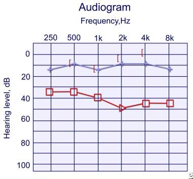
Click to see larger pictureClick to see detailView Full Size Image
Media type:  Graph

Media file 3:  Audiogram 1 year later of the same child as in Image 2. Her functioning in kindergarten continues to be excellent, and she has noticed hearing improvement at home. Her physical examination shows a pinpoint opening at the external auditory canal (EAC) now, and this audiogram confirms improved hearing in the right ear compared to the prior audiogram in Image 2. Her right conductive hearing loss is shown by the distance between the black brackets and the red triangles.
Click to see larger pictureClick to see detailView Full Size Image
Media type:  Graph

Media file 4:  CT scan in the axial plane of right unilateral external auditory canal atresia (EACA) is shown. The right external auditory canal (EAC), to the left of this photo, is not developed compared to the normally developed left EAC. The image is slightly tilted in the horizontal plane.
Click to see larger pictureClick to see detailView Full Size Image
Media type:  CT

Media file 5:  CT scan showing unilateral auditory canal atresia in coronal section of the right ear. No ear canal is seen to the left of this picture (patient's right).
Click to see larger pictureClick to see detailView Full Size Image
Media type:  CT


  1. Jahrsdoerfer, RA. Congenital Atresia of the Ear. Laryngoscope. 1978;88(9 pt. 3 Suppl 13):1-48. [Medline][Full Text].
  2. Caughey RJ, Jahrsdoerfer RA, Kesser BW. Congenital cholesteatoma in a case of congenital aural atresia. Otol Neurotol. Oct 2006;27(7):934-6. [Medline].
  3. Sheykholeslami K, Sheykholesami K, Megerian CA, Kaga K. Vestibular-evoked myogenic potentials in infancy and early childhood. Laryngoscope. Aug 2005;115(8):1440-4. [Medline].
  4. Ishimoto S, Ito K, Karino S, Takegoshi H, Kaga K, Yamasoba T. Hearing levels in patients with microtia: correlation with temporal bone malformation. Laryngoscope. Mar 2007;117(3):461-5. [Medline].
  5. Sheykholeslami K, Mohammad HK, Sebastien S, Kaga K. Binaural interaction of bone-conducted auditory brainstem responses in children with congenital atresia of the external auditory canal. Int J Pediatr Otorhinolaryngol. 2003;67(10):1083-90. [Medline].
  6. Siegert R, Mattheis S, Kasic J. Fully implantable hearing aids in patients with congenital auricular atresia. Laryngoscope. Feb 2007;117(2):336-40. [Medline].
  7. Lambert PR. Major congenital ear malformations: surgical management and results. Ann Otol Rhinol Laryngol. Nov-Dec 1988;97(6 Pt 1):641-9. [Medline].
  8. Jahrsdoerfer RA, Yeakley JW, Aguilar EA, et al. Grading system for the selection of patients with congenital aural atresia. Am J Otol. Jan 1992;13(1):6-12. [Medline].
  9. Glasscock ME 3rd, Schwaber MK, Nissen AJ, Jackson CG. Management of congenital ear malformations. Ann Otol Rhinol Laryngol. Sep-Oct 1983;92(5 Pt 1):504-9. [Medline].
  10. Battelino S, Hocevar-Boltezar I, Zargi M. Intraoperative use of mitomycin C in fibrous atresia of the external auditory canal. Ear Nose Throat J. Dec 2005;84(12):776-9. [Medline].
  11. Yamane H, Sunami K, Takayama M, et al. Application of modified transmastoid approach methods to congenital atresia of the external ear canal with middle ear infection. Acta Otolaryngol Suppl. Oct 2004;38-44. [Medline].
  12. Teufert KB, De la Cruz A. Advances in congenital aural atresia surgery: effects on outcome. Otolaryngol Head Neck Surg. 2004;131(3):263-70. [Medline].
  13. Satar B, Yetiser S, Ozkaptan Y. Acoustic characteristics of reconstructed ear canal after atresia surgery. Br J Audiol. 2000;34(6):379-81. [Medline].
  14. De la Cruz A, Linthicum FH Jr, Luxford WM. Congenital atresia of the external auditory canal. Laryngoscope. Apr 1985;95(4):421-7. [Medline].
  15. Wetmore RF, Muntz HR, McGill TJ. Congenital malformations of the ear. In: Pediatric Otolaryngology Principles and Practice Pathways. 2000:235-52.
  16. Evans AK, Kazahaya K. Canal atresia: "surgery or implantable hearing devices? The expert's question is revisited". Int J Pediatr Otorhinolaryngol. Mar 2007;71(3):367-74. [Medline].
  17. Blevins NH, Byahatti SV, Karmody CS. External auditory canal duplication anomalies associated with congenital aural atresia. J Laryngol Otol. Jan 2003;117(1):32-8. [Medline].
  18. Caversaccio M, Romualdez J, Baechler R, et al. Valuable use of computer-aided surgery in congenital bony aural atresia. J Laryngol Otol. Apr 2003;117(4):241-8. [Medline].
  19. Hol MK, Cremers CW, Coppens-Schellekens W, Snik AF. The BAHA Softband. A new treatment for young children with bilateral congenital aural atresia. Int J Pediatr Otorhinolaryngol. Jul 2005;69(7):973-80. [Medline].
  20. Jacobsen N, Mills R. Management of stenosis and acquired atresia of the external auditory meatus. J Laryngol Otol. Apr 2006;120(4):266-71. [Medline].
  21. Parrish KL, Amedee RG. Atresia of the external auditory canal. J La State Med Soc. Sep 1990;142(9):9-12. [Medline].
  22. Swartz JD, Faerber EN. Congenital malformations of the external and middle ear: high-resolution CT findings of surgical import. AJR Am J Roentgenol. Mar 1985;144(3):501-6. [Medline].

External Auditory Canal Atresia excerpt

Article Last Updated: Jul 18, 2008