Practice Essentials
Achondrogenesis is characterized by severe micromelia, macrocrania, and short trunk. Although rare, it is the second most common lethal skeletal dysplasia after thanatophoric dysplasia
Achondrogenesis is classified as type I (ACGIA, ACGIB) and type II forms (ACGII). Achondrogenesis type II, hypochondrogenesis, and neonatal spondyloepiphyseal dysplasia congenita are now known to be phenotypic variants of the same disorder.
Achondrogenesis results from mutations in the TRIP11 (ACGIA), SLC26A2 (ACGIB), and COL2A1 (ACGII) genes, with achondrogenesis types IA and IB being autosomal recessive conditions and achondrogenesis type II most often being sporadic. Prenatally, polyhydramnios and hydrops leading to fetal demise can be observed. Skeletal anomalies include severe micromelia, a short and narrow thorax with pulmonary hypoplasia, and absent or very abnormal ossification of the skull and vertebral bodies.
Background
Marco Fraccaro first described achondrogenesis in 1952. [1] He used the term to describe a stillborn female with severe micromelia and marked histologic cartilage changes. The term was later used to characterize the most severe forms of chondrodysplasia in humans, which were invariably lethal before or shortly after birth. By the 1970s, researchers concluded that achondrogenesis was a heterogeneous group of chondrodysplasias lethal to neonates; achondrogenesis type I (Fraccaro-Houston-Harris type) and type II (Langer-Saldino type) were distinguished on the basis of radiologic and histologic criteria.
See the image below.
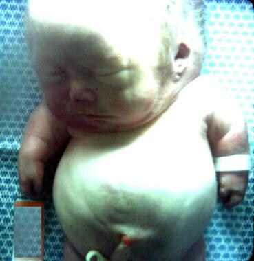 An infant with achondrogenesis type II. Note the disproportionately large head, large and prominent forehead, flat facial plane, flat nasal bridge, small nose with severely anteverted nostrils, micrognathia, extremely short neck, short and flared thorax, protuberant abdomen, and extremely short upper extremities.
In 1983, a new radiologic classification of achondrogenesis (types I-IV) by Whitley and Gorlin was adopted in the McKusick catalog. [2] According to this classification, type I and type II have the same femoral cylinder index (CIfemur; calculated as length of femur divided by width of femur) range (1-2.8). Both types have crenated ilia and stellate long bones. Multiple rib fractures are characteristic of type I but not type II. Type III has nonfractured ribs, halberd ilia, mushroom-stem long bones, and a CIfemur of 2.8-4.9. Type IV has nonfractured ribs, sculpted ilia, well-developed long bones, and a CIfemur of 4.9-8. This radiologic classification based on the CIfemur was later abandoned. Researchers suggested that achondrogenesis type III probably corresponds to type II and that type IV probably corresponds to mild type II (hypochondrogenesis).
See the images below.
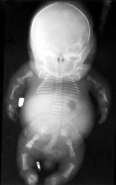 This posteroanterior (PA) view radiograph of an infant with achondrogenesis type II shows the relatively large calvaria with normal cranial ossification, short and flared thorax, bell-shaped cage and shorter ribs without fractures, relatively well ossified iliac bone with long crescent-shaped medial and inferior margins, and short tubular bones. The sacrum, pubis, and ischium are not visible.
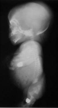 Lateral view radiograph of an infant with achondrogenesis type II. Note the relatively large head with a normal cranial ossification and enlarged fontanelles, short ribs, absent sternal ossification, ossification only in anterior parts of the vertebral bodies, and short and curved femora.
In the late 1980s, structural mutations in collagen II were shown to cause achondrogenesis type II, which thus constitutes the severe end of the spectrum of collagen II chondrodysplasias. Achondrogenesis type I was subdivided further in 1988 on the basis of convincing histologic criteria. It was subdivided into type IA, which has apparently normal cartilage matrix but inclusions in chondrocytes, and type IB, which has an abnormal cartilage matrix. Classification of type IB as a separate group has been confirmed by the discovery of its association with mutations in the diastrophic dysplasia sulfate transporter (DDST) gene, making it allelic with diastrophic dysplasia.
Currently, three variants of achondrogenesis have been defined based on radiologic and histopathologic features: type IA (Houston-Harris), type IB (Parenti-Fraccaro), and type II (Langer-Saldino). Achondrogenesis IA is caused by recessive mutations in the TRIP11 gene, type IB results from recessive mutations of the SLC26A2 gene, and type II is caused by autosomal dominant mutations of the COL2A1 gene.
Achondrogenesis II results from heterozygosity for a new dominant mutation in the COL2A1 gene [3] at the chromosomal locus 12q8.11–q13.2. Intramolecular heterogeneity has been recognized, and genotype–phenotype correlations have been demonstrated. [4]
Several heritable osteochondrodysplasias have now been recognized as members of the family of type II collagen disorders, all of which result from dominant mutations in the COL2A1 gene. [5, 6, 4] Phenotypes within this group range from severe lethal dwarfism at birth to relatively mild conditions with precocious osteoarthrosis and little or no skeletal growth abnormality. Achondrogenesis II-hypochondrogenesis and lethal spondyloepiphyseal dysplasia congenita (SEDC) represent the more severe end of the spectrum. [7] These entities are characterized by severe disproportionate short stature of prenatal onset. The distinction between these phenotypes is mainly based on clinical, radiographic, and morphological features but considerable phenotypic overlap often hampers proper classification.
Mutations within the COL2A1 gene also cause hypochondrogenesis (OMIM 200610), spondyloepiphyseal dysplasia (SED) congenita (OMIM 183900), SED Namaqualand type (OMIM 142670), mild SED with precocious osteoarthritis, spondyloepimetaphyseal dysplasia Strudwick type (OMIM 184250), Kniest dysplasia (OMIM 156550), multiple epiphyseal dysplasia with myopia and conductive deafness, spondyloperipheral dysplasia (OMIM 271700), and Stickler dysplasia type I (OMIM 108300). [8, 9]
Pathophysiology
Achondrogenesis type IA (ACGIA; OMIM 200600):
Mutation of the TRIP11 gene, located on chromosome 14q32, has been identified in ACGIA. Mutational change leads to abnormal secretion of Golgi microtubule-associated protein 210 (GMAP-210), a protein associated with skeletal development.
Achondrogenesis type IB (ACGIB; OMIM 600972):
A series of mutations in the SLC26A2 gene, on chromosome 5q32, have been identified in patients with ACGIB. Homozygosity or compound heterozygosity for these mutations leads to premature stop codons or structural mutations in transmembrane domains and abnormal synthesis of sulfated proteoglycans in cartilage.
Achondrogenesis type II (ACGII; OMIN 256050):
Heterozygous mutations in the COL2A1 gene, located on chromosome 12q13, have been identified in ACGII, as well as in other type II collagenopathies (eg, spondyloepiphyseal dysplasias, hypochondrogenesis). Type II has a single base change, substituting serine for glycine in the type II procollagen gene of the alpha 1(II) chain. This disrupts the triple helix formation, leading to a paucity of type II collagen in the cartilage matrix. Epiphyseal cartilage lacks type II collagen. It is replaced by type I and type III collagens, which are not normally produced by chondrocytes. Differentiated chondrocytes do not express type II collagen. In addition to skeletal abnormalities, severe pulmonary hypoplasia, thought to be directly related to the underlying pathology in collagen expression, is associated with achondrogenesis.
Type II achondrogenesis/hypochondrogenesis (Whitley and Gorlin prototype IV) has immunohistologic findings that demonstrate apparent abnormal intracellular accumulation of type II collagen within vacuolar structures of chondrocytes. This suggests the presence of abnormal, poorly secreted type II collagen. Molecular defects of type II collagen and new dominant mutations account for the observed phenotype.
Epidemiology
Frequency
United States
Lethal achondrogenesis types I and II are both rare, with prevalence ranging from 0.09-0.23 in 10,000 births.
Mortality/Morbidity
Achondrogenesis type I results in stillbirth more frequently than type II. Babies with achondrogenesis type I who are not stillborn typically have a shorter gestation and survive for a shorter time than those with type II. They are also smaller with much shorter limbs, which supports the general view that type I is the more severe form.
Race
Achondrogenesis has no racial predilection.
Sex
Males and females are equally affected.
Age
Achondrogenesis is detected prenatally or at birth because of typical clinical, radiologic, histologic, and molecular findings.
-
An infant with achondrogenesis type II. Note the disproportionately large head, large and prominent forehead, flat facial plane, flat nasal bridge, small nose with severely anteverted nostrils, micrognathia, extremely short neck, short and flared thorax, protuberant abdomen, and extremely short upper extremities.
-
This posteroanterior (PA) view radiograph of an infant with achondrogenesis type II shows the relatively large calvaria with normal cranial ossification, short and flared thorax, bell-shaped cage and shorter ribs without fractures, relatively well ossified iliac bone with long crescent-shaped medial and inferior margins, and short tubular bones. The sacrum, pubis, and ischium are not visible.
-
Lateral view radiograph of an infant with achondrogenesis type II. Note the relatively large head with a normal cranial ossification and enlarged fontanelles, short ribs, absent sternal ossification, ossification only in anterior parts of the vertebral bodies, and short and curved femora.
-
An infant with achondrogenesis type II. Note the protuberant abdomen and extremely short lower extremities.
-
Photomicrographs of the costal cartilage of an infant with achondrogenesis type II. This shows prominent hypercellularity, large chondrocytes, deficient matrix, and abnormally large, stellate cartilage canals. The left image is X42, and the right image is X106.
Tables
| IA | IB | II | |
|---|---|---|---|
| Skull | Absent or severely reduced ossification | Normal or reduced ossification for age | Normal ossification |
| Long bones | Short and bowed Stellate, longitudinally oriented Metaphyseal spurring |
Very short, square or stellate Metaphyseal spurring Poor ossification of phalanges |
Short and bowed Metaphyseal flaring and cupping |
| Thorax | Short, barrel-shaped Ribs: Short and horizontally oriented, with splayed anterior ends Multiple fractures (beaded appearance) |
Ribs: Short and thin, typically no fractures | Short, barrel- or bell-shaped Short ribs, no fractures |
| Spine | Vertebral bodies: Absent or rudimentary ossification |
Vertebral bodies: Absent or rudimentary central ossification Vertebral lateral pedicles: Usually ossified |
Variable Vertebral bodies and pedicles: Ossified or unossified |
| Pelvis | Sacrum: Absent or rudimentary ossification |
Iliac bone: Abnormal ossification giving a crescent-shaped appearance Ischium: Not ossified. |
Halberd-like iliac bone Ischial and pubic bones: Unossified |


