Background
Ptosis, also referred to as blepharoptosis, is defined as an abnormal low-lying upper eyelid margin with the eye in primary gaze. The normal adult upper lid lies 1.5 mm below the superior corneal limbus and is highest just nasal to the pupil. [1] See the images below.
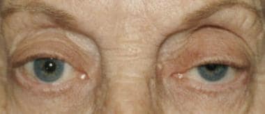 Left ptosis. Lid crease is absent on the left. The crease is up in the sulcus. Superior sulcus deformity is present on the left and right, and the patient is elevating her brows. The right upper lid should be checked for an underlying or masked ptosis. If the right lid is ptotic, lifting the left lid causes the right lid to droop.
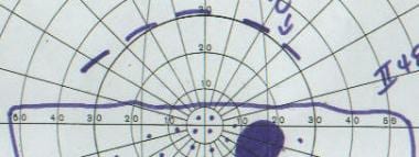 Visual field shows functional blockage of superior visual field due to a ptotic lid. Hashed line represents the superior extent of the seen visual field with the lid lifted. Solid line is with the lid in its natural, ptotic position.
Ptosis can be classified as congenital, as shown below, or acquired. [2] This differentiation is based on age. A more comprehensive classification is based on etiology and includes myogenic, aponeurotic, neurogenic, mechanical, traumatic, and pseudoptotic. The most common cause of congenital ptosis is myogenic due to the improper development of the levator muscle. [3]
Most cases of acquired ptosis are secondary to aponeurotic causes, such as involutional changes, a disinsertion, or a dehiscence. Identification of the underlying pathophysiologic mechanism is paramount to institute proper treatment.
Pathophysiology
Ptosis is the result of dysfunctioning of one or both upper eyelid elevator muscles. These elevator muscles are the levator palpebrae superioris and the Mueller muscle.
The levator palpebrae superioris is a striated muscle innervated by the superior division of the oculomotor nerve (cranial nerve III). This muscle is about 40 mm long and originates from the lesser wing of the sphenoid. It continues anteriorly, and at the Whitnall ligament, it travels inferiorly as an aponeurosis. The aponeurosis is 14-20 mm long and inserts into the anterior aspect of the tarsal plate. It also sends attachments to the skin, forming the upper eyelid crease. The levator muscle and aponeurosis is the major elevator of the upper eyelid.
The Mueller muscle, a sympathetically innervated smooth muscle, originates from the undersurface of the levator superioris. Approximately 12 mm long, it inserts superiorly on the tarsal border and elevates the upper eyelid by approximately 2 mm.
Epidemiology
Mortality/Morbidity
Mortality associated with ptosis usually results from anesthetic complications from surgery. Kearns-Sayre disease, a subtype of chronic progressive external ophthalmoplegia, is a syndrome with associated myogenic ptosis, retinal pigmentary changes, and cardiac conduction abnormalities that can cause death.
Morbidity is associated with blockage of the visual axis in the severely ptotic eyelid. Congenital cases can obstruct vision and lead to amblyopia. Even without visual axis obstruction, the eyelid may induce refractive errors, especially astigmatism resulting in amblyopia.
In adults, the morbidity is associated with constriction of the superior visual fields. Patients may complain that they tire easily when reading and experience frontal headaches as they lift their eyebrows in an effort to keep the eyelids open. Patients may be dissatisfied with their appearance.
Race
No racial predilection has been described for ptosis.
Sex
No sexual predilection has been described for ptosis.
Age
Acquired ptosis can occur at any age, but it is commonly seen in older adults. Congenital ptosis occurs at birth.
Patient Education
Inform patients that symmetry is difficult, if not impossible, to achieve (see Medical/Legal Pitfalls).
-
Patient with bilateral ptosis before surgery. Note the high lid creases.
-
Same patient as in the previous image after bilateral internal levator advancement. No skin incision was made, and no crease reformation was performed.
-
Anterior approach to the levator. White band is the levator aponeurosis (arrow).
-
Left ptosis. Lid crease is absent on the left. The crease is up in the sulcus. Superior sulcus deformity is present on the left and right, and the patient is elevating her brows. The right upper lid should be checked for an underlying or masked ptosis. If the right lid is ptotic, lifting the left lid causes the right lid to droop.
-
Visual field shows functional blockage of superior visual field due to a ptotic lid. Hashed line represents the superior extent of the seen visual field with the lid lifted. Solid line is with the lid in its natural, ptotic position.
-
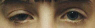
Congenital ptosis on right. Note the presence of a lid crease.
-
Glasses with a crutch attached (arrow) that can be used to lift the lid if the patient does not desire surgery.
-
Patient with myasthenia gravis. Right lid is more ptotic than the left lid.
-
Same patient as in the previous image, 3 months later. Note how the ptosis has changed and is more on the left than the right.
-
Patient with bilateral ptosis before surgery.
-
Same patient as in the previous image after internal levator advancement. Patient has excessive skin (dermatochalasia) after the lid was lifted, with a pseudoptotic effect more on the left than the right. The dermatochalasia was present before surgery but is more significant afterward. Patient also has brow ptosis.


